
CARDIAC DIAGNOSTICS
Both cardiac computed tomography (CT) and magnetic resonance imaging (MRI) are fast non-invasive imaging techniques that can save lives by early detection of coronary artery disease and structural disease of the heart itself.
Coronary artery disease develops, when the blood vessels that supply the heart with oxygen and nutrients develop deposits of fatty substances within the vessel wall. These deposits are called plaque. When plaques grow over time, the vessels are narrowed and blood flow to the heart decreases. resulting in symptoms like chest pain or shortness of breath. A complete blockage of the vessel causes a heart attack.
The amount of plaque and the growth rate are influenced by many factors such as age, sex, tobacco use, high blood pressure, obesity or a family history of coronary artery disease.

CT Coronary Angiography
Coronary artery (left anterior descending artery) with a non calcified plaque (arrow) with significant narrowing of the lumen.

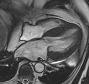
Heart MRI
Longitudinal section of the heart (2-chamber view): The bright tissue (arrow) is scar tissue after a myocardial infarction.
Heart MRI
Markedly thickened myocrdium (heart muscle) in a patient with a cardiomyopathy (hypertrophic cardiomyopathy).
Modern imaging technologies such as Computed tomography coronary angiography (CCTA) can detect plaques before they cause symptoms. As plaques calcify over time CT can be used to quantify calcifications and to guide the medical therapy for patients with a high risk for coronary artery disease. In patients with mild symptoms CCTA can also be used to rule out coronary artery disease as the cause of symptoms. However, patients with an acute onset of chest pain should present to an emergency room as soon as possible.
Computed tomography uses radiation to image the human anatomy, but with modern scanners, the radiation exposure of a typical scan remains below the annual radiation exposure from natural sources. In contrast, magnetic resonance tomography (MRI) uses no radiation for imaging the human body. Cardiac MRI can be used to assess cardiac function and to detect scar tissue in the heart muscle (myocardium) after a heart attack. Also called myocardial infarction. MRI allows to assess the amount of tissue damage after an infarction and helps to decide whether a revascularization procedure should be performed to reestablish blood flow to the myocardium affected by the infarct.
Cardiac MRI is also helpful to detect diseases of the myocardium itself or inflammatory diseases such as myocarditis.